
An echocardiogram is an ultrasound imaging procedure that uses high-frequency sound waves and a computer to create images of the heart. It may be done while the patient is lying down, sitting up, and even walking around. Echocardiograms can show whether your heart chambers and valves are enlarged or abnormally shaped. They can also show fluid in one or both of your lungs. The images produced by echocardiography can be printed on film or stored as digital images on a CD-ROM.
A Port Saint Lucie echocardiogram specialist can recommend the procedure if your healthcare provider suspects you have a heart problem.
Table of Contents
How Is It Done?
In a cardiac sonographer’s office, you will be asked to remove your shirt and any objects that might interfere with the test, such as jewelry or a belt. You also may need to wear a gown. You’ll lie on an examination table that has a large “cushion” over it. The cushion keeps you from feeling pressure on your chest. You’ll be asked to hold your breath for short periods, or you may have a tube placed in your nose that releases air as you breathe out. This test allows the sound waves to record clearly inside your heart.
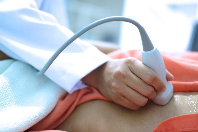
An echocardiogram is done by placing a small transducer device onto your skin. The transducer sends and receives sound waves. It is connected to a computer by a cable. The machine’s sound waves bounce off your heart, and the computer turns them into moving pictures of your heart on a monitor or printout.
What Does it Show?
The pictures made by an echocardiogram show the size, shape, movement, and function of your heart. An echocardiogram can help you identify conditions such as abnormal heart valves, which may not close properly or stick together. This abnormality can cause blood to back up toward your lungs or make it hard for your heart to pump enough blood. It can also help detect whether your heart is working harder than it should or if you have more blood than usual in your body.
There are many different echocardiograms, depending on what the doctor wants to look at. If you need an echocardiogram, your healthcare provider will explain what type is right for you.
How to Prepare
You don’t need to do anything special to prepare for an echocardiogram, although you may be asked not to eat or drink for at least four hours before the test.
The echocardiogram specialist will let you know if there are any restrictions on what you can or cannot eat or drink before your procedure since certain foods and drinks can affect the test results.
How It Feels
You may feel some pressure on your chest as the transducer is placed over your skin. You may feel a little bit of pain or discomfort during deep breathing, when you hold your breath, or when air is released into your system through a tube placed in your nose. The examination is painless, and it should not take longer than 30 minutes.
In summary, an echocardiogram is an ultrasound imaging procedure that uses high-frequency sound waves and a computer to create images of the heart. It is done by placing a transducer on your skin which sends and receives soundwaves that translate them into images. An echocardiogram shows the size, shape, movement, and function of your heart. Your doctor will give you instructions on how to prepare for the procedure. You should not experience any pain during the process.